Understanding the Physics of Intense X-ray Interactions
X-ray free-electron lasers (XFELs) make it possible to image biological particles, such as viruses and macromolecules with extremely intense bursts of X-rays. Any sample exposed to a single pulse of such high energy will be strongly ionized and thus severely damaged. The key to obtaining a clear picture or diffraction pattern is to outrun radiation damage, by using shorter bursts of x-ray light than the time needed for these damage processes to evolve. The aim for this research is to understand the relation between structural damage of biological samples induced by short intense pulses from XFELs, and the possibilities of solving a structure from a perturbed diffraction pattern.
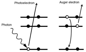
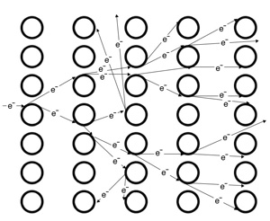
One of the major damage generators, in a biological sample zapped by XFEL, is the secondary ionizations induced by the ejected electrons (as illustrated by figure 1) colliding with bound electrons causing secondary ionization. The number of secondary electrons generated from a single photo ionization event, can be more than an order of magnitude higher than the direct ionizations (figure 2). To model and describe the damage of a protein in an XFEL pulse, it is important to understand the ionization dynamics. We mainly work with two independent theoretical models to try to understand the damage processes. One model is based on the Ionizing Molecular Dynamics (IMD) approach, and the other is the Non-Local Thermal Equilibrium (non-LTE) plasma approach.

Figure 1
The secondary electron ejection. Auger decay is one of two principal processes for the relaxation of an inner shell electron vacancy in an excited or ionized atom. The Auger effect is a two electron process in which an electron makes a discrete transition from a higher shell to fill a vacant inner shell hole. The energy gained in this process is transferred to another bound electron which then escapes from the atom.
The photoelectron and the Auger electron are released at different times, and have different energies. The figure on the left shows a high energy photoelectron leaving the inner shell. The figure on the right shows the release of the low energy Auger electron as the system relaxes. Auger decay is the dominant relaxation process for filling K-hole vacancies in light elements, like C, N, O, S, P.

Figure 2
Secondary electron cascades. The initial electron, emitted through the Auger decay or direct photo ionization, leads to an avalanche of electrons due to inelastic scattering.
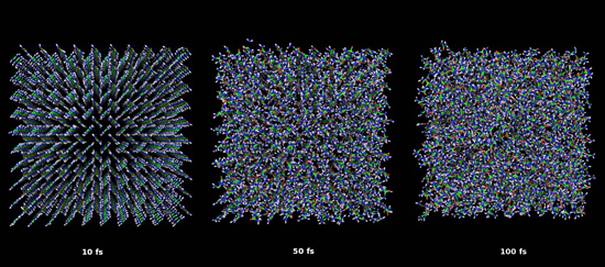
The IMD model is based on electron impact ionization cross sections in a solid sample. Those are calculated using a combination of first principle quantum calculations of the energy loss function of a material and an empirical model for calculating the cross sections of interest. Once the cross sections are determined we run stochastic simulations of the secondary electron generation based on a single initial electron with a certain energy. The next step is to utilize these ionization rates in the molecular dynamics code, to model the ionization due to an external x-ray pulse and to calculate the subsequent effect on the atomic positions. With this approach we can follow trajectory and scattering strength of each atom and hence calculate the diffraction pattern that is expected to be recorded from the same pulse. An illustration of an IMD simulation is shown in figure 3.

Figure 3
Ultrafast damage dynamics of urea nanocrystal. The structure of 10x10x10 unit cells of crystalline urea (simulated using the IMD model with periodic boundary conditions) after exposure to an X-ray pulse at 160 J/cm2 with different pulse lengths: left - 10 fs (FWHM), middle - 50 fs and right - 100 fs.
The second model is based on a non-LTE plasma code, including radiation transfer, which follows the atomic populations in time through an average ion model. The rates for all processes are calculated and a self-consistent rate-equation is solved that determines the evolution of the atomic populations. This is referred to as Collision-Radiative Equation which is the standard method to describe the population kinetics. The model is very accurate once the ionization in the sample has reached a certain degree.
Until very recently the theoretical predictions were rather difficult to compare to experiments, since XFEL sources were not available. Since the opening of the XFEL source at Stanford, LCLS, to users in fall 2009, the tools necessary to confirm or disapprove our theoretical models are now available. Using data from our experiments, we aim to develop better models that will tell us more about the relation between matter and short intense x-ray pulses, and in particular increase our understanding of the ionization dynamics caused by radiation damage. The interplay between experiments and theory is crucial to understand the extreme conditions of matter in an XFEL experiment. The modeling is used to guide molecular imaging and protein nanocrystals experiments and the experimental results from known samples are used to verify and develop the models.